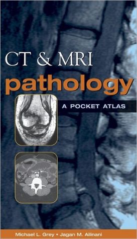
CT & MRI Pathology: A Pocket Atlas
This pocket atlas includes unmatched state-of-the-art CT and MRI images of 110 common pathologies organized by body system and type of pathology. The easy-reference format provides a concise overview of pathology, etiology, epidemiology, signs and symptoms, imaging characteristics, treatment, and prognosis. Features a separate pediatric section and detailed index guides.
Search in google:
This new, one-of-a-kind portable resource offers fast access to high-quality images covering 110 common pathologies visualized on CT and MRI. CT & MRI Pathology is the perfect tool for an overview of the pathology, etiology, epidemiology, signs & symptoms, imaging characteristics, treatment, and prognosis.EXCELLENT REFERENCE IMAGESDISEASE SUMMARIESQUICK LOOKUPSPORTABILITY*Hundreds of images for on-the-spot comparisons*Capsule summaries put pathology, etiology, epidemiology, imaging characteristics, treatment, and prognosis at your fingertips *Organization by body system and pathologies enhance learning*Extensive, detailed index guides you quickly to what you wantMichael L.Grey, MS RT(R)(MR)(CT) is certified in MR and CT and teaches in the radiologic technology program at Southern Illinois University, Carbondale, Illinois.Jagan Mohan Ailinani, MD is Clinical Professor of Radiology, Southern Illinois University School of Medicine, Carbondale, Illinois, and visiting faculty, St. Louis University School of Medicine, St. Louis, Missouri. He is Diplomat of American Board of Radiology. Doody Review Services Reviewer:Cris Anne Zimmermann, R.T.(R)(Froedtert Hospital)Description:This MRI and CT pocket atlas of common pathology provides a wealth of images along with clinical data to demonstrate the specific pathology.Purpose:The purpose of this book is to provide a handy tool for the technologist or student in CT and MRI. The book contains only common pathology seen on a day-to-day basis in an imaging facility. The author meets the objectives and the handbook provides a concise view of common pathology through text and images. This book is needed for the professional CT and MRI technologist who wants to stay abreast of common pathological anomalies.Audience:The book is simply written for students and technologists in CT and MRI. Features:The book covers common pathology seen in CT and MR images. The central nervous system, head and neck, chest and mediastinum, abdomen, pelvis and musculoskeletal systems are all included in this concise handbook. This book divides pathology, in each section, by congenital, neoplasms, trauma, and vascular diseases and infectious anomalies. The unique features, such as a list of etiology, signs and symptoms, treatment and prognosis, are especially helpful for one trying to gain insight into a disease. Another nice feature are the arrows marking the pathology on the images, along with a brief description of what they are highlighting.Assessment:For an imaging professional, this book would be a nice tool. Since most pocket books are atlases of normal anatomy, it's nice to see a pocketbook that covers common pathology in a concise manner. Everything from acoustic neuromas topneumothoraces to rotator cuff tears are covered and are more common in an imaging facility than normal anatomy. This book provides insight into the disease along with useful images depicting the pathology.
\ From The CriticsReviewer: Cris A. Zimmermann, RT(R)(Froedtert Hospital)\ Description: This MRI and CT pocket atlas of common pathology provides a wealth of images along with clinical data to demonstrate the specific pathology.\ Purpose: The purpose of this book is to provide a handy tool for the technologist or student in CT and MRI. The book contains only common pathology seen on a day-to-day basis in an imaging facility. The author meets the objectives and the handbook provides a concise view of common pathology through text and images. This book is needed for the professional CT and MRI technologist who wants to stay abreast of common pathological anomalies.\ Audience: The book is simply written for students and technologists in CT and MRI. \ Features: The book covers common pathology seen in CT and MR images. The central nervous system, head and neck, chest and mediastinum, abdomen, pelvis and musculoskeletal systems are all included in this concise handbook. This book divides pathology, in each section, by congenital, neoplasms, trauma, and vascular diseases and infectious anomalies. The unique features, such as a list of etiology, signs and symptoms, treatment and prognosis, are especially helpful for one trying to gain insight into a disease. Another nice feature are the arrows marking the pathology on the images, along with a brief description of what they are highlighting.\ Assessment: For an imaging professional, this book would be a nice tool. Since most pocket books are atlases of normal anatomy, it's nice to see a pocketbook that covers common pathology in a concise manner. Everything from acoustic neuromas to pneumothoraces to rotator cuff tears are covered and are more common in an imaging facility than normal anatomy. This book provides insight into the disease along with useful images depicting the pathology.\ \ \ 3 Stars from Doody\ \
