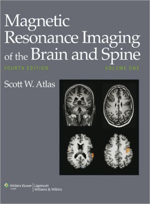
Magnetic Resonance Imaging of the Brain and Spine (Two Volume Set)
Established as the leading textbook on imaging diagnosis of brain and spine disorders, Magnetic Resonance Imaging of the Brain and Spine is now in its Fourth Edition. This thoroughly updated two-volume reference delivers cutting-edge information on nearly every aspect of clinical neuroradiology. Expert neuroradiologists, innovative renowned MRI physicists, and experienced leading clinical neurospecialists from all over the world show how to generate state-of-the-art images and define...
Search in google:
Established as the leading textbook on imaging diagnosis of brain and spine disorders, Magnetic Resonance Imaging of the Brain and Spine is now in its Fourth Edition. This thoroughly updated two-volume reference delivers cutting-edge information on nearly every aspect of clinical neuroradiology. Expert neuroradiologists, innovative renowned MRI physicists, and experienced leading clinical neurospecialists from all over the world show how to generate state-of-the-art images and define diagnoses from crucial clinical/pathologic MR imaging correlations for neurologic, neurosurgical, and psychiatric diseases spanning fetal CNS anomalies to disorders of the aging brain. Highlights of this edition include over 6,800 images of remarkable quality, more color images, and new information using advanced techniques, including perfusion and diffusion MRI and functional MRI.A companion Website will offer the fully searchable text and an image bank. Stephen C. Voron The second edition of this book consists of 32 chapters: 15 on the brain, including the skull base, temporal bone, and orbit; 6 on the spine; and 11 primarily on physics. Compared with the first edition, published in 1991, the chapters on the temporal bone and on functional MRI of the brain are new, and other chapters have been extensively revised and expanded. The editor has fulfilled his goal of improving upon the definitive work on MRI of central nervous system diseases. This field has continued to grow during the past five years, and this up-to-date revision is of true value. Dr. Atlas has assembled 57 contributors, all experts and educators in their fields, including physicists, neuropathologists, neurologists, neurosurgeons, and neuroradiologists, to compile a comprehensive book on the science as well as the technical and clinical aspects of MRI. As a result, this book should appeal to radiologists, neuroradiologists, neurologists, and neurosurgeons as well as to neuroscience personnel needing a clinical reference. The book is well illustrated, including MRI scans of very good quality, colored artist's renditions, colored histopathology and, new to this revision, colored pathology specimens. Each chapter begins with a chapter outline and ends with an extensive reference list. This revision is a commendable update of a classic text. In addition to new features mentioned above, it includes more than 500 new pages and is well worth purchasing by owners of the first edition. At $245.00, the book is reasonably priced for an extensively illustrated radiology work. I recommend the book to trainees and experienced individuals in the appropriate specialties, hospital and departmentlibraries, and bookstores.
Contributing AuthorsPreface to the First EditionPrefaceAcknowledgmentsPt. IPrinciples1Instrumentation: Magnets, Coils, and Hardware32Contrast Development and Manipulation in MR Imaging333Principles of Image Formation594Contrast Agents and Relaxation Effects795Fundamentals of Flow and Hemodynamics1016Fast Imaging Principles1277Diffusion and Diffusion Tensor MR Imaging1978Perfusion MR Imaging2159Artifacts in MR239Pt. IIBrain10Disorders of Brain Development27911Central Nervous System Manifestations of the Phakomatoses and Other Inherited Syndromes37112Epilepsy41513White Matter Diseases and Inherited Metabolic Disorders45714Intraaxial Brain Tumors56515Extraaxial Brain Tumors69516Intracranial Hemorrhage77317Intracranial Vascular Malformations and Aneurysms83318Cerebral Ischemia and Infarction91919MR Angiography: Techniques and Clinical Applications98120Head Trauma105921Intracranial Infection109922Normal Aging, Dementia, and Neurodegenerative Disease1177Pt. IIISkull Base23The Skull Base124324The Sella Turcica and Parasellar Region128325Anatomy and Diseases of the Temporal Bone136326Eye, Orbit, and Visual System1433Pt. IVSpine and Spinal Cord27Congenital Anomalies of the Spine and Spinal Cord: Embryology and Malformations152728Degenerative Disease of the Spine163329Neoplastic Disease of the Spine and Spinal Cord171530Spinal Trauma176931Vascular Disorders of the Spine and Spinal Cord182532Spinal Infection and Inflammatory Disorders1855Pt. VAdvanced Applications33Clinical Functional MR Imaging197334Psychiatric Disease199335MR Spectroscopy and the Biochemical Basis of Neurological Disese2021Subject Index
\ Reviewer: James Milburn, MD(Ochsner Clinic Foundation)\ Description: This is the fourth edition of a popular textbook on neuroradiology which includes detailed study of the brain, skull base, and spine. The third edition was published in 2002.\ Purpose: It is intended to reinforce proven neuroimaging findings, illustrate the pathologic basis for those findings, and explore the use of emerging and newer MRI methods in the context of contributing significantly to diagnosis and guiding therapy. This fine work clearly meets these are worthy objectives.\ Audience: The audience includes all students of neuroradiology at every level of training, from novice to expert. It is also intended to be useful to all physicians who treat or have an interest in neurologic and spine disorders, including neurologists, neurosurgeons, orthopedic surgeons, neonatologists, and psychiatrists.\ Features: The brain imaging section comprises most of the first volume and is quite complete. A very nice initial section covers the technical aspects and physics of image acquisition. The second volume consists of the skull base, spine and spinal cord, and advanced applications. The advanced imaging applications are up to date at the time of publication and are a significant addition to this fourth edition. The book is well referenced with a very helpful index and the images and diagrams are outstanding. It is well written by many notable experts in the field of neuroradiology.\ Assessment: This book compares favorably to the other complete neuroradiology textbooks on the market. It is written is paragraph format, in contrast to another excellent popular series of textbooks in radiology which are in outline format with bullet points. Personal preference will determine which book readers will use. A new edition is certainly justified, because although neuroradiology is now a mature field, it continues to advance with many new directions and applications.\ \ \ \ \ Stephen C. VoronThe second edition of this book consists of 32 chapters: 15 on the brain, including the skull base, temporal bone, and orbit; 6 on the spine; and 11 primarily on physics. Compared with the first edition, published in 1991, the chapters on the temporal bone and on functional MRI of the brain are new, and other chapters have been extensively revised and expanded. The editor has fulfilled his goal of improving upon the definitive work on MRI of central nervous system diseases. This field has continued to grow during the past five years, and this up-to-date revision is of true value. Dr. Atlas has assembled 57 contributors, all experts and educators in their fields, including physicists, neuropathologists, neurologists, neurosurgeons, and neuroradiologists, to compile a comprehensive book on the science as well as the technical and clinical aspects of MRI. As a result, this book should appeal to radiologists, neuroradiologists, neurologists, and neurosurgeons as well as to neuroscience personnel needing a clinical reference. The book is well illustrated, including MRI scans of very good quality, colored artist's renditions, colored histopathology and, new to this revision, colored pathology specimens. Each chapter begins with a chapter outline and ends with an extensive reference list. This revision is a commendable update of a classic text. In addition to new features mentioned above, it includes more than 500 new pages and is well worth purchasing by owners of the first edition. At $245.00, the book is reasonably priced for an extensively illustrated radiology work. I recommend the book to trainees and experienced individuals in the appropriate specialties, hospital and departmentlibraries, and bookstores.\ \ \ A comprehensive text-reference on MRI of the central nervous system that presents the science as well as the current technical and clinical aspects, for neuroradiologists, neurologists, neurosurgeons, and clinicians who interact to discuss the results of MRI in patients with neurologic disease. Among the changes in this revised and updated edition are the addition of new chapters which encompass not only new technology (such as functional MRI), but also new and potential clinical applications based on recently described clinicopathologic MRI correlations (e.g., diseases of the temporal bone). Thoroughly illustrated, the ample use of color specimens is a new feature in this edition for the purpose of presenting characteristics of lesions that are or may be discernible in advanced MR techniques. Annotation c. Book News, Inc., Portland, OR (booknews.com)\ \ \ \ \ 5 Stars! from Doody\ \


