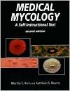
Medical Mycology: A Self-Instructional Text
Each of the seven modules includes prerequisites, content outline, objectives, follow-up activities, references, and self-study examinations Teaches proper laboratory practice and presents the biology and physiology of fungi, descri\ \ The book contains predominantly black-and-white illustrations, with some color illustrations.\
Search in google:
Written for the beginning mycologist, this book discusses the general and specific characteristics, cultivation, and identification of medically important fungi. This highly accessible and user-friendly text is ideal for both self-directed study and classroom instruction. The book contains numerous line drawings and halftone illustrations, as well as 75 full-color plates. The reader is further aided by the listings (in the front and back of the book) of the techniques and procedures presented in the text, the charts in each module, and the color plates. This book features an extensive illustrated glossary. Roberta B. Carey This is a second edition of a self-instructional text of medical mycology originally published in 1985. Although the number of pages has not changed, the methods, taxonomy, and terminology have been updated. The workbook is designed to teach medical mycology, the identification of yeasts and molds that cause human infections. This is a difficult discipline for students to learn because identification is based on observation rather than objective test results. This manual covers the basic terms essential to discriminate between species of fungi. It supplements exposure to mycology in a clinical lab setting or a classroom. Written for the beginning mycologist or medical technology student, the authors have applied their extensive knowledge to help the inexperienced appreciate the details of structure. The facts are presented in easy-to-learn modules, which make the material less awesome to comprehend. Almost every page has black-and-white photographs and companion drawings to emphasize the structures. Color plates demonstrate the color and texture of the colonies. The size of each photograph (2 by 3 inches) is adequate to see the growth characteristics of each fungus. There is an excellent glossary at the end of the book, and as new terms are introduced in a chapter they are presented in bold font. Procedures and media formulations are highlighted separately in boxes for easy reference. The first edition of this text has been essential in teaching mycology and as a quick resource for terminology; the second edition will continue this tradition. The combination of information, illustrations, and exam questions with answers provides a comprehensive learning package. The text isnot designed to cover all aspects of fungal infections. However, reading this text is an important first step in acquiring knowledge about organisms that cause human infections and how to identify them.
List of Techniques and ProceduresList of ChartsList of Color PlatesIntroduction: How to Use This TextModule 1Basics of MycologyModule 2Laboratory Procedures for Fungal Culture and IsolationModule 3Common Fungal OpportunistsModule 4Superficial and Dermatophytic FungiModule 5YeastsModule 6Organisms Causing Subcutaneous MycosesModule 7Organisms Causing Systemic MycosesApp. A: Answers for Study Questions and Final ExamsApp. B: Common SynonymsApp. C: GlossaryApp. D: List of ManufacturersColor PlatesIndex
\ From The CriticsReviewer: Roberta B. Carey, PhD(Loyola University Medical Center)\ Description: This is a second edition of a self-instructional text of medical mycology originally published in 1985. Although the number of pages has not changed, the methods, taxonomy, and terminology have been updated.\ Purpose: The workbook is designed to teach medical mycology, the identification of yeasts and molds that cause human infections. This is a difficult discipline for students to learn because identification is based on observation rather than objective test results. This manual covers the basic terms essential to discriminate between species of fungi. It supplements exposure to mycology in a clinical lab setting or a classroom.\ Audience: Written for the beginning mycologist or medical technology student, the authors have applied their extensive knowledge to help the inexperienced appreciate the details of structure. The facts are presented in easy-to-learn modules, which make the material less awesome to comprehend.\ Features: Almost every page has black-and-white photographs and companion drawings to emphasize the structures. Color plates demonstrate the color and texture of the colonies. The size of each photograph (2 by 3 inches) is adequate to see the growth characteristics of each fungus. There is an excellent glossary at the end of the book, and as new terms are introduced in a chapter they are presented in bold font. Procedures and media formulations are highlighted separately in boxes for easy reference.\ Assessment: The first edition of this text has been essential in teaching mycology and as a quick resource for terminology; the second edition will continue this tradition. The combination of information, illustrations, and exam questions with answers provides a comprehensive learning package. The text is not designed to cover all aspects of fungal infections. However, reading this text is an important first step in acquiring knowledge about organisms that cause human infections and how to identify them.\ \ \ \ \ Roberta B. CareyThis is a second edition of a self-instructional text of medical mycology originally published in 1985. Although the number of pages has not changed, the methods, taxonomy, and terminology have been updated. The workbook is designed to teach medical mycology, the identification of yeasts and molds that cause human infections. This is a difficult discipline for students to learn because identification is based on observation rather than objective test results. This manual covers the basic terms essential to discriminate between species of fungi. It supplements exposure to mycology in a clinical lab setting or a classroom. Written for the beginning mycologist or medical technology student, the authors have applied their extensive knowledge to help the inexperienced appreciate the details of structure. The facts are presented in easy-to-learn modules, which make the material less awesome to comprehend. Almost every page has black-and-white photographs and companion drawings to emphasize the structures. Color plates demonstrate the color and texture of the colonies. The size of each photograph (2 by 3 inches) is adequate to see the growth characteristics of each fungus. There is an excellent glossary at the end of the book, and as new terms are introduced in a chapter they are presented in bold font. Procedures and media formulations are highlighted separately in boxes for easy reference. The first edition of this text has been essential in teaching mycology and as a quick resource for terminology; the second edition will continue this tradition. The combination of information, illustrations, and exam questions with answers provides a comprehensive learning package. The text isnot designed to cover all aspects of fungal infections. However, reading this text is an important first step in acquiring knowledge about organisms that cause human infections and how to identify them.\ \ \ 3 Stars from Doody\ \
