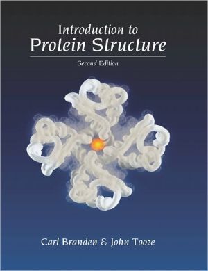
Introduction to Protein Structure
Introduction to Protein Structure provides an account of the principles of protein structure, with examples of key proteins in their biological context generously illustrated in full-color to illuminate the structural principles described in the text. The first few chapters introduce the general principles of protein structure both for novices and for non-specialists needing a primer. Subsequent chapters use specific examples of proteins to show how they fulfill a wide variety of biological...
Search in google:
Introduction to Protein Structure provides an account of the principles of protein structure, with examples of key proteins in their biological context generously illustrated in full-color to illuminate the structural principles described in the text. The first few chapters introduce the general principles of protein structure both for novices and for non-specialists needing a primer. Subsequent chapters use specific examples of proteins to show how they fulfill a wide variety of biological functions. The book ends with chapters on the experimental approach to determining and predicting protein structure, as well as engineering new proteins to modify their functions. Candace J. Krepel This text provides an excellent overview of protein structure and how structure and function interrelate. The previous edition was published in 1991. The purpose is to convey an understanding of protein structure as a basis for the understanding of biological reactions. The text is written at a content level consistent with advanced undergraduates or above. A basic knowledge of proteins and their biological function is assumed. Basic protein structure (primary, secondary, etc.) is reviewed, followed by elucidation of the structures of examples from various protein families (that is, proteins with common functions), and to demonstrate how function follows form. The editors use excellent interrelated illustrations that allow the reader to understand the three-dimensional aspects of the structures. Amino acid sequence is listed, secondary structure of alpha helixes and beta sheets are shown, then three-dimensional models and drawings demonstrate the tertiary structure. The editors use these to show how the proteins intersect and interact with the biological environment. They do an excellent job of showing proteins as being an important part of the living organism. The text offers an up-to-date look at what is known about protein structure and the methodologies for determining structure. The editors use well-chosen paradigms as examples. In this rapidly growing field, this new edition is welcome.
The x-ray structure of DNA complexes with 434 Cro and repressor revealed novel features of protein-DNA interactions\ The general features of the model for DNA binding were confirmed experimentally in 1987 when Stephen Harrison's group at Harvard University determined the structure of a complex of DNA and the DNA-binding domain of the 434 repressor to 3.2 A resolution. However, it also became evident, both from the structure of this complex and from further site-directed mutagenesis studies, that the selective recognition of the different operator regions by the 434 repressor depends mostly on other factors than the amino acid residues of the recognition helix. The complexity of the fine tuning of DNA regulation has been clearly demonstrated by Harrison's subsequent studies of complexes between different operator DNA regions and both 434 Cro and the DNA-binding domain of 434 repressor.\ For purely practical reasons, the complexes that Harrison first studied contained the N-terminal DNA-binding domain of the repressor from phage 434, which comprises 69 amino acids, complexed with a 14 base-pair piece of synthetic DNA (" 14mer") having a completely palindromic sequence. In other words, the DNA in the complex had a strict twofold symmetry analogous to the twofold symmetry of the dimeric repressor molecule. This synthetic DNA thus contains identical halves, each of which, as we shall see, binds one subunit of Cro or one repressor fragment. The three 14 base-pair operator regions that the 434 repressor recognizes in the phage right-hand operator (OR) are not perfectly palindromic, however (Table 8.2). The 14mer is closest in sequence to a site (OL2) in a second operator region of the phage, the "left" operator (OL), as Table 8.2 shows. The only difference is an inversion of base pair 7 from A-T to T-A, and experiments had shown that this inversion did not alter the affinity for the DNA of either intact repressor or the N-terminal DNA-binding fragment.\ The crystals of the complex with 14mer diffracted to only medium resolution, however, and by systematic variations of the length of the DNA fragment and its sequences at the ends, Harrison and coworkers were later able to find a piece of DNA that gave crystals that diffracted to high resolution, both with 434 Cro and the DNA-binding domain of 434 repressor. This DNA fragment contains 20 nucleotides in each chain, and the sequence of its middle region is identical to OR1 (see Table 8.2). The 5' ends contain one nonpaired nucleotide that is involved in packing the fragments in the crystal.\ By comparing the crystal structures of these complexes with a further complex of the 434 repressor DNA-binding domain and a synthetic DNA containing the operator region OR3, Harrison has been able to resolve at least in part the structural basis for the differential binding affinity of 434 Cro and repressor to the different 434 operator regions.\ The structures of 434 Cro and the 434 repressor DNAbinding domain are very similar\ The 434 Cro molecule contains 71 amino acid residues that show 48% sequence identity to the 69 residues that form the N-terminal DNA-binding domain of 434 repressor. It is not surprising, therefore, that their threedimensional structures are very similar (Figure 8.11). The main difference lies in two extra amino acids at the N-terminus of the Cro molecule. These are not involved in the function of Cro. By choosing the 434 Cro and repressor molecules for his studies, Harrison eliminated the possibility that any gross structural difference of these two molecules can account for their different DNA-binding properties. The DNA-binding domain of 434 repressor also has significant sequence homology (26% identity) with the corresponding part of the lambda repressor and, consequently, a related three-dimensional structure (compare Figures 8.7 and 8.11). Like its lambda counterpart, the subunit structure of the DNAbinding domain of 434 repressor, as well as that of 434 Cro, contains a cluster of four a helices, with helices 2 and 3 forming the helix-turn-helix motif. The two helix-turn-helix motifs are at either end of the dimer and contribute the main protein-DNA interactions, while protein-protein interactions at the C-terminal part of the chains hold the two subunits together in the complexes. Both 434 Cro and repressor fragments are monomers in solution even at high protein concentrations, whereas they form dimers when they are bound to DNA. (It should be noted once again, however, that in the intact repressor the main dimerization interactions are believed to be formed by the C-terminal domain, which is not present in the crystal complex.).....
PART 1 BASIC STRUCTURAL PRINCIPLES 1. The Building Blocks 2. Motifs of Protein Structure 3. Alpha-Domain Structures 4. Alpha/Beta Structures 5. Beta Structures 6. Folding and Flexibility 7. DNA Structures PART 2 STRUCTURE, FUNCTION AND ENGINEERING 8. DNA Recognition in Procaryotes by Helix-Turn-Helix Motifs 9. DNA Recognition by Eukaryotic Transcription Factors 10. Specific Transcription Factors Belong to a Few Families 11. An Example of Enzyme Catalysis: Serine Proteinases 12. Membrane Proteins 13. Signal Transduction 14. Fibrous Proteins 15. Recognition of Foreign Molecules by the Immune System 16. The Structure of Spherical Viruses 17. Prediction, Engineering, and Design of Protein Structures 18. Determination of Protein Structures
\ From The CriticsReviewer: Candace J. Krepel, MS(Medical College of Wisconsin)\ Description: This text provides an excellent overview of protein structure and how structure and function interrelate. The previous edition was published in 1991.\ Purpose: The purpose is to convey an understanding of protein structure as a basis for the understanding of biological reactions. \ Audience: The text is written at a content level consistent with advanced undergraduates or above. A basic knowledge of proteins and their biological function is assumed.\ Features: Basic protein structure (primary, secondary, etc.) is reviewed, followed by elucidation of the structures of examples from various protein families (that is, proteins with common functions), and to demonstrate how function follows form. The editors use excellent interrelated illustrations that allow the reader to understand the three-dimensional aspects of the structures. Amino acid sequence is listed, secondary structure of alpha helixes and beta sheets are shown, then three-dimensional models and drawings demonstrate the tertiary structure. The editors use these to show how the proteins intersect and interact with the biological environment. They do an excellent job of showing proteins as being an important part of the living organism.\ Assessment: The text offers an up-to-date look at what is known about protein structure and the methodologies for determining structure. The editors use well-chosen paradigms as examples. In this rapidly growing field, this new edition is welcome.\ \ \ \ \ Candace J. KrepelThis text provides an excellent overview of protein structure and how structure and function interrelate. The previous edition was published in 1991. The purpose is to convey an understanding of protein structure as a basis for the understanding of biological reactions. The text is written at a content level consistent with advanced undergraduates or above. A basic knowledge of proteins and their biological function is assumed. Basic protein structure (primary, secondary, etc.) is reviewed, followed by elucidation of the structures of examples from various protein families (that is, proteins with common functions), and to demonstrate how function follows form. The editors use excellent interrelated illustrations that allow the reader to understand the three-dimensional aspects of the structures. Amino acid sequence is listed, secondary structure of alpha helixes and beta sheets are shown, then three-dimensional models and drawings demonstrate the tertiary structure. The editors use these to show how the proteins intersect and interact with the biological environment. They do an excellent job of showing proteins as being an important part of the living organism. The text offers an up-to-date look at what is known about protein structure and the methodologies for determining structure. The editors use well-chosen paradigms as examples. In this rapidly growing field, this new edition is welcome.\ \ \ BooknewsIllustrates (in clear color diagrams) and explains for both biologists and chemists the structural and functional logic that has emerged from the latest accumulated data on protein structures. Annotation c. Book News, Inc., Portland, OR (booknews.com)\ \ \ \ \ 5 Stars! from Doody\ \


